Visual Plasticity
The visual system is an excellent model for exploring how the quality of experiences guides the function of developing brain circuitry. The lab employs several complementary techniques to study plasticity within the circuitry of the visual system.
In vivo calcium imaging of neuronal activity in visual cortex of alert mice
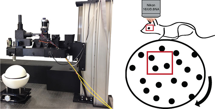
On the left, the Neurolabware microscope for in vivo imaging. Head-fixed mice are imaged atop a floating styrofoam ball. The position of the ball is tracked, as it the area of the pupil. These metrics reveal the relative attentiveness of the mouse during presentation of a visual stimulus.
An example of imaging neuronal activity in alert mice during a visual stimulus repeated three times. The activity of three neurons transduced with AAV:pSyn-GCaMP6s (circled in red, green and blue) are presented during these three consecutive presentations of the movie. The traces in the corresponding colors at left correspond to each neuron.
Multi-unit electrophysiology

This is the classic method of measuring and comparing the responses of multiple neurons (units) to a visual stimulus presented to each eye individually. In this example, we have presented a series of gratings at different orientations to one eye and measured the local change in potential with resin-coated tungsten microelectrodes.
Optical Imaging of Intrinsic signals

Optical imaging of intrinsic signals (OIS) measures the reflectance of light off the surface of the brain. The signal correlates with local brain acitivty. We employ the method of measuring the magnitude of the OIS signal to a periodic stimulus developed by Kalatsky and Stryker at UCSF. This technique is non-invasive and provides good spatial resolution of the cortical region responsive to a visual (or somatosensory) stimulus. We often use OIS to map the functional region corresponding to sensory cortex for two-photon in vivo imaging of dendritic spines and axonal boutons
Behavioural metrics of visual acuity (The visual water task)
")
The visual water task is a two-choice water maze in which the position of a hidden platform indicated by a monitor presenting a sinusoidal spatial frequency grating rather than an isoluminant grey screen. Mice readily build this association and we use this test to estimate their visual acuity by determining the spatial frequency at which they can no longer discriminate the grating from the blank screen